








Ophthalmology

Home

Speciality

Ophthalmology

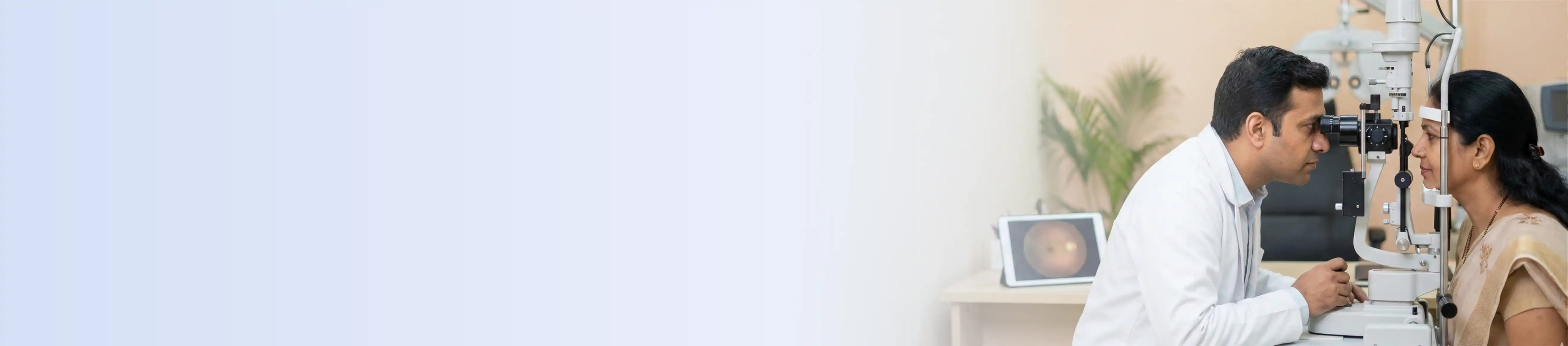
The Department of Ophthalmology at Saraswathi Institute of Medical Sciences has been treating patients with eye conditions since 2008. When it first opened, the setup was simple a basic OPD and one operating theatre. Over the years, the department has grown into a proper eye care centre that now handles everything from routine vision check-ups to complicated ocular surgeries requiring microsurgical precision.

The range of conditions treated here is wide. Patients come in with cataracts, glaucoma, retinal diseases, corneal disorders, paediatric eye problems, eye injuries, and conditions needing oculoplastic surgery. Depending on what the patient needs, treatment can be medical, surgical, or both. The department does not push one approach the clinical team looks at what the patient actually needs and plans accordingly.

In 2021, the department took a significant step forward by launching a formal MS Ophthalmology postgraduate training program. This turned the department into a recognized teaching and training centre, not just a place where patients are treated, but a place where the next generation of eye specialists is being shaped. Residents here get real surgical exposure, structured academic sessions, and research opportunities that go beyond textbook learning.

The underlying commitment of the department has not changed since day one deliver accessible, affordable, and high-quality eye care to patients from all walks of life and do whatever is clinically possible to reduce preventable blindness in the community.

Key Differentiators

Handling Complex Eye Conditions

Some eye conditions are straightforward. Others carry a genuine risk of permanent vision loss if not caught early or managed properly. The department has built experience in handling difficult cases diabetic retinopathy, retinal detachment, uveitis, age-related macular degeneration, corneal perforations, and pediatric retinal disorders. Getting to these conditions early, before they cross a point of no return, is what the clinical team focuses on.
 

Ocular Trauma (Emergency Care)

Eye injuries cannot wait. When someone comes in with a corneal laceration, scleral injury, chemical eye injury, eyelid trauma, or penetrating ocular trauma, fast and accurate management is what protects the eye. The department handles these emergencies with the surgical skills and equipment needed to give the patient the best possible chance of keeping their vision.
 

Eye and Systemic Disease 

The eye often shows the first visible signs of diseases happening elsewhere in the body. Patients with diabetes mellitus, hypertension, autoimmune disorders, and neurological conditions frequently develop eye complications that, if caught early, can be managed before they cause serious damage. The department works closely with other specialties to make sure these patients get regular retinal screening and timely ophthalmic intervention as part of their overall care.
 

Minimally Invasive Surgical Approach

Wherever possible, the department uses less invasive techniques, cause less trauma to the eye, and allow faster recovery. Phacoemulsification cataract surgery, for example, is done through a tiny incision the eye heals faster, the patient gets back to normal vision sooner, and the risk of complications is lower compared to older techniques.
 

Advanced Technologies

Optical Coherence Tomography (OCT) - Huvitz

High-resolution cross-sectional images of the retinal layers and structures of the optic nerve are provided. Essential for the diagnosis of macular disease, monitoring glaucoma, and monitoring retinal disease over time.

Automated Perimetry - Cyrix Healthcare

Describes the patient’s visual field. It is often used to assess glaucoma and neuro-ophthalmic disorders that cause loss of peripheral vision.

Appasamy Associates

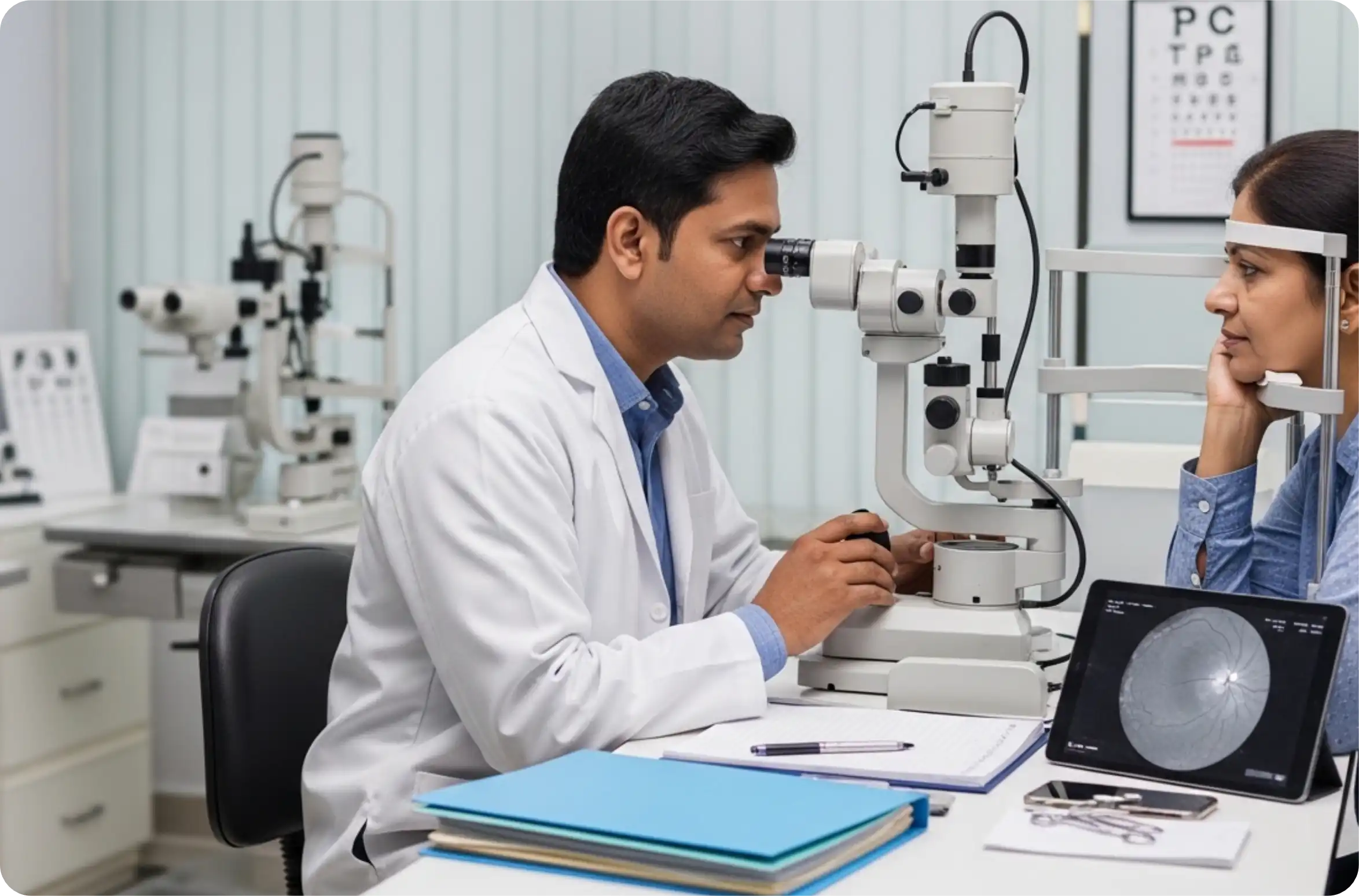
Slit Lamp Units (5 Units) Five slit lamp stations allow detailed examination of the anterior segment, cornea, lens, iris and anterior chamber with high magnification.

Autorefractometer & Keratometer - Nidek

Accurate measurement of refractive errors and corneal curvature. Helps prescribe glasses and plan corneal procedures.

Non-Contact Tonometer - Nidek

Measures Intraocular Pressure Without Touching The Eye, An Important Step In Glaucoma Screening And Monitoring

A-Scan Biometry App - Appasamy 

Associates calculate the correct intraocular lens (IOL) power before cataract surgery. Accurate biometry directly affects how well the patient sees after the operation.

Retinal Examination Tools

The department uses a Heine direct ophthalmoscope, Heine streak retinoscope, indirect ophthalmoscope, and Volk 90D and 20D lenses, giving the clinical team a thorough view of the posterior segment and retina during examination.

Procedures

The department performs phacoemulsification cataract surgery as the primary method for cataract removal, a technique that uses ultrasound energy to break up the cloudy lens through a small incision. Small incision cataract surgery is also performed when required. Postoperative procedures, like YAG laser posterior capsulotomy and laser peripheral iridotomy are usually carried out in the laser room, and not really requiring a formal operation, or at least not in the usual sense.
 

Glaucoma is a condition where the optic nerve gets damaged, usually silently, without the patient noticing until significant vision is already lost. The department manages it through regular intraocular pressure monitoring, visual field testing, medical therapy, trabeculectomy when surgery is needed, and laser procedures. Catching it early is the only way to prevent permanent vision loss.
 

The retina unit manages diabetic retinopathy, applies retinal laser therapy, uses OCT imaging to track disease progression, and screens premature babies for retinopathy of prematurity (ROP). For diabetic patients especially, regular retinal screening is not optional. It is what stands between them and serious vision loss that could have been avoided.
 

Procedures performed include dacryocystorhinostomy (DCR) for blocked tear ducts, dacryocystectomy (DCT), ptosis correction for drooping eyelids, entropion and ectropion repair, eyelid tumour excision with reconstruction, and skin grafting where needed. These procedures restore both function and appearance around the eye area.

The department manages corneal laceration repair, scleral injury repair, eyelid laceration repair, corneal perforation treatment, bandage contact lens application, and tissue adhesive (glue) therapy for cases where suturing is not the right option. Emergency trauma cases are taken up quickly in ocular trauma, every hour matters

Misaligned eyes whether in a child or an adult, affect how the visual system develops and how the person is perceived socially. The department corrects paralytic squint, restrictive strabismus, and paediatric squint disorders surgically, improving binocular vision and eye alignment.

Additional services include pterygium excision with conjunctival autograft, evisceration, enucleation, syringing and probing for blocked tear ducts in infants, and routine minor ophthalmic procedures handled in OPD.
 

The MS Ophthalmology postgraduate training program has been running since 2021 and has added real academic weight to the department. Residents get hands-on surgical training, exposure to a wide mix of ophthalmic conditions, and regular academic sessions, including seminars, journal clubs, case presentations, and surgical demonstrations.
Research focus areas currently include tissue adhesive therapy, corneal healing, advanced surgical techniques, and retinal disease management. The department runs on the principle that good clinical practice and good research go hand in hand one feeds into the other.

Join our newsletter today to get the latest updates and easily manage your data more efficiently!
Newsletter Subscribe