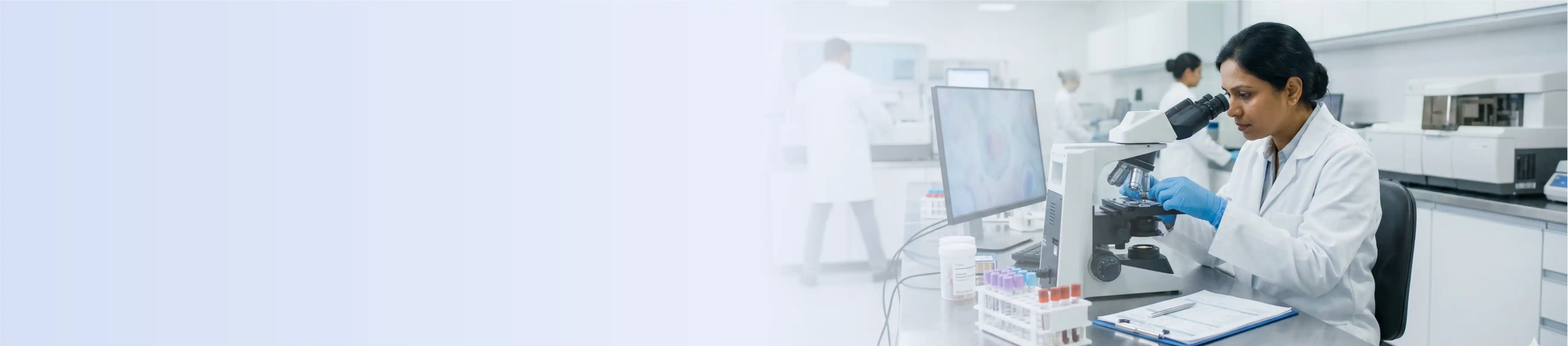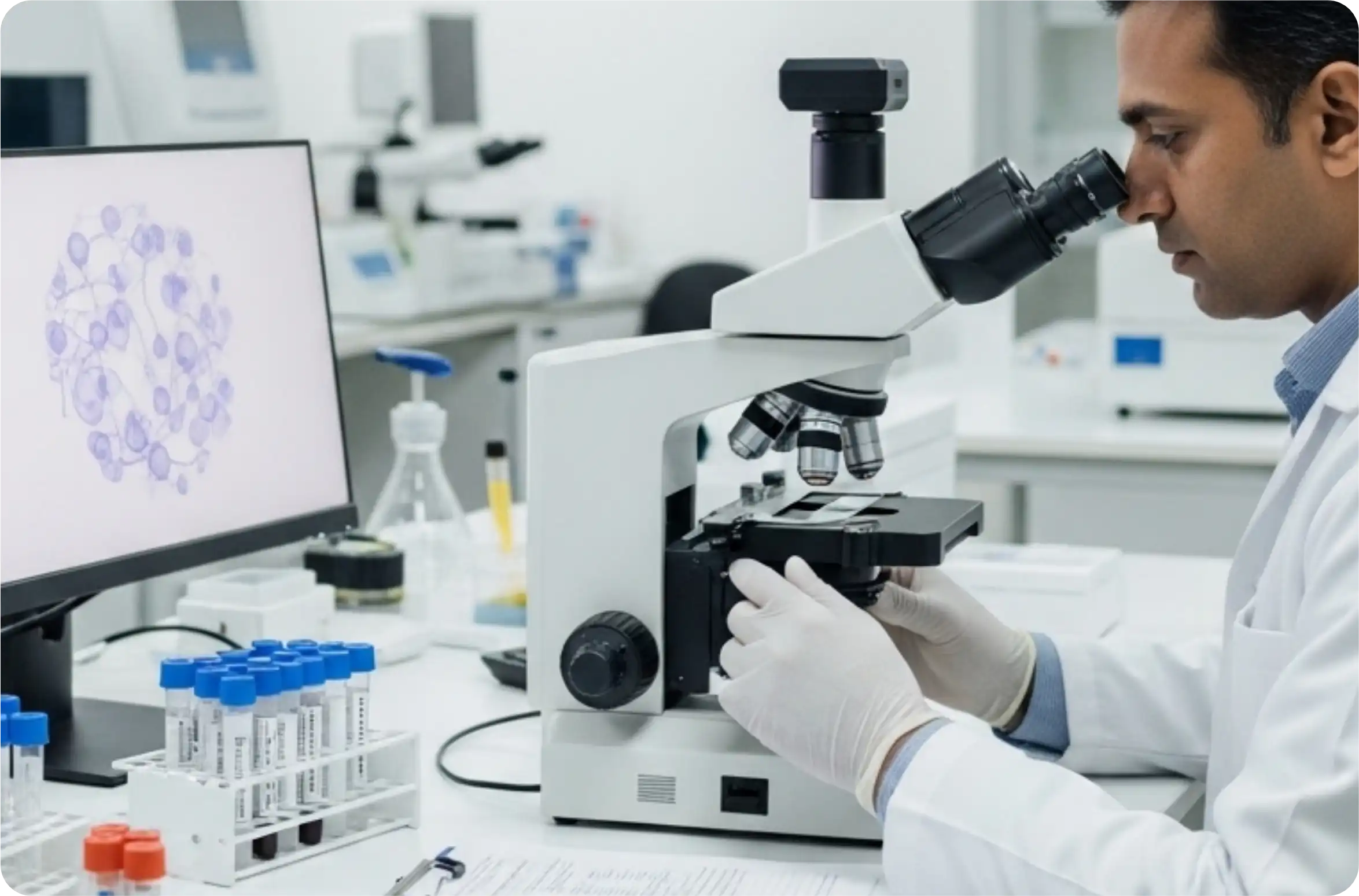
The Department of Pathology is one of the most important pillars of modern healthcare to help clinicians with accurate laboratory diagnosis, disease monitoring and prognostic evaluation. The department uses cutting-edge diagnostic technology and scientific accuracy to provide accurate and timely reports that help guide patient treatment decisions across multiple specialties.
The department integrates histopathology, cytology, hematology and clinical pathology services under one roof to offer comprehensive diagnostic support for routine and complex medical conditions. The department is highly committed to quality assurance, rapid turnaround time and academic excellence and makes significant contribution to patient care, medical education and clinical research.
Histology: The histopathology section provides detailed tissue based diagnosis by microscopic examination of biopsy and surgical specimens. Each specimen is treated with standardized protocols to maintain tissue architecture and cellular detail for accurate interpretation.
The department routinely performs special staining techniques, immunohistochemistry (IHC), to help in tumour typing, grading, staging and prognostic assessment. These advanced diagnostic modalities help in differentiating benign and malignant conditions and help clinicians in formulating appropriate treatment strategies. Histopathology services are essential for oncology, gastrointestinal diseases, gynaecological disorders, renal diseases and inflammatory conditions.
Cytology : The Cytology unit is involved in the early diagnosis of diseases, especially those that are minimally invasive, using cellular examination techniques. The department provides Fine Needle Aspiration Cytology (FNAC), exfoliative cytology and body fluid cytology for prompt diagnostic evaluation. FNAC procedures are fast and economical in evaluation of swellings, lymph nodes, thyroid lesions, breast lumps and soft tissue masses.
Exfoliative cytology including Pap smear examination is useful in screening and early detection of pre-cancerous and cancerous lesions. The cytology section lays emphasis on accuracy, patient comfort and prompt reporting for early medical intervention.
Haematology: Management of various hematological and systemic diseases. The section has an automated haematology analyser which can perform complete blood counts, reticulocyte counts, peripheral smear analysis, and coagulation studies with high precision.
The department is also important in diagnosing anemia, leukemias, infections, platelet disorders, clotting abnormalities and other blood related diseases. The diagnostic accuracy is further improved by peripheral smear examination by experienced pathologists who can recognize abnormal cellular morphology. HbA1c testing is also available for long term monitoring of diabetes mellitus to help in effective glycemic control and patient management.
Clinical Pathology: The Clinical Pathology section offers a wide range of laboratory investigations for diagnosis, disease monitoring and therapeutics assessment. The laboratory performs routine and special examination of urine, semen, cerebrospinal fluid, synovial fluid, pleural fluid, peritoneal fluid and pericardial fluid.
Urinalysis can be used to diagnose urinary tract infections, kidney disorders, metabolic abnormalities, and systemic diseases. Semen analysis helps to evaluate male infertility by assessing sperm count, motility, morphology and seminal parameters . Analysis of body fluids is useful in the diagnosis of infections, inflammatory disorders, malignancies and joint diseases. It differentiates transudates from exudates and helps in the early detection of pathological changes.
The section plays an important role in rapid clinical decision-making through reliable and clinically relevant diagnostic information.
Key Differentiators
Advanced Technologies
Automated Tissue Processing Systems
Tissue specimens undergo a fully automated processing cycle that standardises every stage from dehydration to paraffin embedding, ensuring that slide quality remains consistent regardless of specimen volume. This removes the variability that manual processing introduces and preserves cellular detail precisely as it needs to be for reliable microscopic reading.
Immunohistochemistry (IHC) Platform
The IHC platform applies targeted antibodies directly to tissue sections, revealing protein expression patterns that routine staining simply cannot show. This makes it the method of choice for tumour classification, receptor status assessment, and distinguishing primary from metastatic malignancies.
Special Staining Techniques
Beyond standard H&E staining, the department uses a broad panel of histochemical stains, each selected for a specific diagnostic question, whether it is identifying fungal organisms, detecting amyloid deposits, assessing fibrosis, or characterising glycogen storage. The right stain applied to the right specimen adds a layer of diagnostic certainty that changes treatment decisions.
Automated Haematology Analysers
Blood samples are processed through high-speed automated analysers that generate precise counts and differentials within minutes, with built-in flagging for abnormal results that require pathologist review. Every flagged sample goes to peripheral smear examination, where trained pathologists assess cellular morphology directly before a final report is issued.
HbA1c Testing System (HPLC)
HPLC-based HbA1c measurement separates haemoglobin variants with a level of accuracy that point-of-care devices cannot consistently match, making it the preferred method for monitoring long-term glycaemic control in diabetic patients. Results from this system provide clinicians with a reliable three-month glucose average on which to base treatment adjustments.
Light Microscopy with High-Resolution Imaging
Every diagnostic section, histopathology, cytology, and haematology relies on high-resolution light microscopy as its primary interpretive tool, and the quality of that microscopy directly determines what can and cannot be seen in a specimen. High-resolution optics make subtle morphological changes visible at the cellular level, which is where many diagnoses are ultimately made or missed.
Laboratory Information System (LIS)
From the moment a sample is received to the point a report reaches the clinician, every step is tracked and documented within the LIS, eliminating transcription errors, preventing sample mix-ups, and creating a complete audit trail for every case. Turnaround times are monitored automatically, and critical values trigger immediate notification to the treating team.
Procedures
Histopathologic Examinations
Histopathological examination is the detailed microscopic examination of biopsies and surgical specimens for the diagnosis of inflammatory, infectious, benign and malignant conditions. The tissue samples are processed, stained and evaluated to give a definitive diagnosis and assess the progress of disease. This is of particular importance in cancer diagnosis and surgical pathology.
Special stains
Routine stains fail to show certain tissue elements, microorganisms and pathological changes, which are demonstrated by special stains. These stains provide increased diagnostic clarity, enabling pathologists to more accurately identify complex disease processes.
Immunohistochemistry (IHC)
Immunohistochemistry is an advanced diagnostic technique for tumour typing, prognostic evaluation and cellular markers. IHC is an important tool in oncology and helps in distinguishing the source of the tumour, classifying the malignancy and deciding on targeted therapy.
Fine Needle Aspiration Cytology (FNAC)
FNAC is an easy, least invasive procedure for rapid diagnosis of lumps, nodules and swellings. FNA is the removal of cellular material from suspicious lesions using a fine needle for microscopic examination. The procedure is safe, rapid, inexpensive and very useful in the evaluation of lesions of the thyroid, breast, lymph nodes and soft tissue.
Cytology exfoliative
Exfoliative cytology involves the study of cells that are shed from body surfaces and fluids, either naturally or by mechanical means. Pap smear testing and other procedures can be used for early detection of cervical abnormalities and pre-cancerous conditions. Cytology of body fluids is also useful in diagnosis of infections and malignancies.
Peripheral Smear Examination and Complete Blood Count
The Complete Blood Count (CBC) and peripheral smear examination are basic hematological investigations that are used to evaluate blood cell parameters and detect abnormal cellular morphology. These tests can help diagnose anaemia, infections, leukaemias, platelet disorders and a number of systemic diseases.
Testing for Coagulation Profile
Coagulation studies evaluate the body’s blood clotting mechanisms and help to diagnose bleeding disorders, thrombotic conditions and coagulation abnormalities. These tests are important both before surgery and in monitoring patients on anticoagulant therapy.
Testing for HbA1c
HbA1c testing is useful for long-term assessment of blood glucose control in diabetic patients by measuring average glucose levels over the previous two to three months. This investigation is important for diabetes diagnosis, monitoring and treatment planning.